
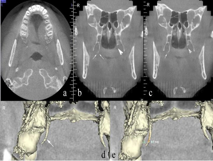
A Cone Beam Computed Tomography (CBCT) study is a valuable tool in dentistry and maxillofacial imaging for assessing various anatomical structures, including the morphology of the pterygoid hamulus. The pterygoid hamulus is a small, hook-like projection of the sphenoid bone located in the posterior aspect of the hard palate, adjacent to the pterygoid process. Its morphology can vary among individuals and can have implications for various dental and surgical procedures.
CBCT imaging offers several advantages over traditional radiographic techniques, providing high-resolution three-dimensional images with minimal distortion. This allows for detailed evaluation of the morphology of the pterygoid hamulus, including its length, width, thickness, and orientation.
In a study involving CBCT evaluation of the pterygoid hamulus morphology, researchers typically recruit a sample of individuals representing a diverse demographic to ensure the findings are applicable across populations. Imaging parameters such as voxel size, field of view, and exposure settings are standardized to ensure consistency across scans.
During the CBCT scan, the patient's head is positioned in the machine, and a series of X-ray images are taken from different angles. These images are then reconstructed using computer algorithms to create a three-dimensional representation of the craniofacial region, including the pterygoid hamulus.
Once the CBCT images are obtained, the morphology of the pterygoid hamulus is analyzed using specialized software. Measurements are taken to assess its dimensions, shape, and any variations in its anatomical features. Common measurements include the length of the hamulus, the angle of its curvature, and its relationship to adjacent structures such as the maxillary tuberosity and the medial pterygoid plate.
The findings of the CBCT study provide valuable information for various clinical applications. For example, knowledge of the morphology of the pterygoid hamulus is essential for clinicians performing procedures such as maxillary tuberosity reduction, orthodontic treatment planning, and surgical interventions in the maxillofacial region. Additionally, understanding variations in the morphology of the pterygoid hamulus can help prevent iatrogenic injuries during procedures involving the soft palate or adjacent structures.
In summary, a Cone Beam Computed Tomography study for the evaluation of the morphology of the pterygoid hamulus utilizes advanced imaging technology to provide detailed three-dimensional information about this anatomical structure. By accurately assessing its dimensions and variations, CBCT imaging enhances treatment planning and improves patient outcomes in various dental and maxillofacial procedures.
No Any Replies to “Cone Beam Computed Tomography Study for Evaluation of Morphology of Pterygoid Hamulus”
Leave a Reply