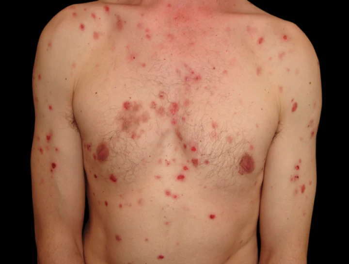
**Pemphigus vulgaris** (PV) is a rare, autoimmune blistering disorder characterized by the formation of painful blisters and erosions on the skin and mucous membranes. The condition arises from the production of autoantibodies that target desmosomal proteins, which are crucial for maintaining cell-to-cell adhesion in epithelial tissues. Here’s a comprehensive overview of pemphigus vulgaris, including its clinical features, diagnosis, and management:
### Clinical Features
**1. **Oral Manifestations:**
- **Initial Symptoms:** Often starts with painful oral mucosal lesions, including ulcers, erosions, and blisters.
- **Lesion Appearance:** Lesions can affect any part of the oral mucosa, including the buccal mucosa, tongue, gingiva, and palate. The blisters are typically fragile and may rupture easily, leading to painful erosions.
- **Chronicity:** Oral lesions can be persistent and challenging to manage, significantly affecting the patient’s quality of life.
**2. **Skin Manifestations:**
- **Blisters and Erosions:** Typically appear on the scalp, face, trunk, and extremities. Blisters may be superficial and prone to rupture, resulting in erosive lesions.
- **Nikolsky Sign:** A positive Nikolsky sign (the skin sloughs off when gently rubbed) is often observed in pemphigus vulgaris.
**3. **Systemic Manifestations:**
- **General Symptoms:** Fever, malaise, and weight loss may occur, especially in severe cases.
- **Complications:** Secondary infections and complications from chronic lesions can arise, impacting overall health.
### Pathophysiology
**1. **Autoimmune Response:**
- **Autoantibodies:** PV is characterized by the presence of autoantibodies directed against desmoglein 1 and desmoglein 3, which are components of desmosomes that mediate cell adhesion in the epidermis and mucous membranes.
- **Acantholysis:** The binding of these autoantibodies disrupts desmosomal connections between keratinocytes, leading to cell separation (acantholysis) and blister formation.
### Diagnosis
**1. **Clinical Examination:**
- **Evaluation of Lesions:** A detailed clinical examination is essential to identify characteristic lesions and assess their distribution.
**2. **Histopathology:**
- **Skin/Mucosal Biopsy:** Biopsy of lesional skin or mucosa is crucial. Histological examination shows acantholysis and blister formation in the epithelial layer.
- **Direct Immunofluorescence:** Demonstrates intercellular deposition of autoantibodies in the epidermis.
**3. **Serology:**
- **Detection of Autoantibodies:** Enzyme-linked immunosorbent assay (ELISA) can detect circulating autoantibodies against desmoglein 1 and 3.
**4. **Indirect Immunofluorescence:**
- **Autoantibody Titers:** Useful in assessing the presence and titer of circulating autoantibodies and monitoring disease activity.
### Management
**1. **Medical Treatment:**
- **Corticosteroids:** High-dose systemic corticosteroids (e.g., prednisone) are the mainstay of treatment to control inflammation and immune response.
- **Immunosuppressive Agents:** Additional immunosuppressive drugs, such as azathioprine, mycophenolate mofetil, or methotrexate, may be used to reduce steroid dependence and manage disease activity.
- **Biologics:** Rituximab, an anti-CD20 monoclonal antibody, is increasingly used in refractory or severe cases, targeting B cells responsible for autoantibody production.
**2. **Supportive Care:**
- **Wound Care:** Proper care of oral and skin lesions is essential to prevent secondary infections and promote healing. This includes maintaining oral hygiene and using topical treatments to soothe lesions.
- **Nutritional Support:** Addressing nutritional deficiencies and providing dietary modifications to manage oral pain and difficulty eating.
**3. **Monitoring and Follow-Up:**
- **Regular Monitoring:** Continuous monitoring of disease activity, treatment efficacy, and potential side effects of medications is crucial.
- **Disease Remission:** Long-term follow-up is necessary to manage and potentially taper down medication as the disease comes under control.
### Prognosis
- **Variable Course:** The disease can range from mild to severe, with the severity of symptoms and response to treatment varying among patients.
- **Chronicity:** PV is often a chronic condition requiring ongoing management, but with effective treatment, many patients can achieve good control of symptoms and an improved quality of life.
### Conclusion
Pemphigus vulgaris is a serious autoimmune blistering disorder that primarily affects the skin and mucous membranes. Early diagnosis and aggressive treatment are crucial for managing the disease and preventing complications. A multidisciplinary approach involving dermatologists, oral surgeons, and other healthcare professionals is essential for comprehensive management, addressing both the systemic and mucosal aspects of the disease.


No Any Replies to “Pemphigus Vulgaris”
Leave a Reply