


The submandibular glands are a pair of glands situated on the floor of the mouth, below the lower jaw. They are one of the three pairs of glands that produce saliva. Submandibular glands can become swollen when small stones block the ducts that supply saliva to the mouth. Sometimes this can lead to an infection.
Anatomy
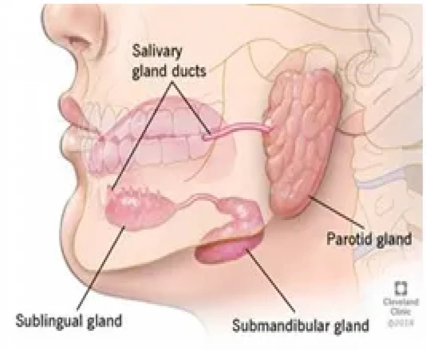
The submandibular glands are the second largest of the three main salivary glands—about the size of a walnut. The two other types of salivary glands are the parotid (the largest) and sublingual glands.
The submandibular glands sit in the submandibular triangle, located underneath the mandible (lower jaw bone) and above the hyoid (tongue) bone. The mylohyoid muscle, a paired muscle that forms the floor of the mouth, separates a superficial and deep lobe in the gland.
The submandibular duct, also called the Wharton's duct, is the excretory duct of the gland. It drains saliva from the glands at the base of the tongue.
The blood supply to the gland comes from the facial artery and lingual artery. The parasympathetic and sympathetic nervous systems stimulate the salivary glands.
Function
The submandibular gland produces saliva, which moistens the mouth and aids in chewing, swallowing, digestion, and helps to keep the mouth and teeth clean. Unstimulated, the submandibular glands provide the majority of saliva to the mouth. On stimulation, the parotid gland takes over, producing the majority of saliva.
The parasympathetic nervous system and the sympathetic nervous system regulate the glands. The parasympathetic system, via the facial nerve, causes the gland to produce secretions and increase blood supply to the gland.
The sympathetic nervous system is responsible for decreasing blood flow and secretions. This results in more enzymes in the saliva, which is essential for digesting food.
Wharton’s Duct
The submandibular duct (also known as Wharton's duct) allows the passage of saliva from the submandibular gland to the sublingual papilla located anteriorly.
The duct extends anteriorly from the submandibular gland superior to the lingual nerve and submandibular ganglion curving over the posterior edge of the mylohyoid muscle into the sublingual space. It is approximately 5-6 cm in length and has a diameter of approximately 1-3 mm on conventional sialographic images.
It runs forward with lingual nerve and vein and hypoglossal nerve and opens into the sublingual papilla at the side of the frenulum of the tongue.
No Any Replies to “Submandibular Glands”
Leave a Reply