A supernumerary tooth is an additional tooth to the normal set of teeth. It may closely resemble the teeth of the group to which it belongs, i.e., molars, premolars or anterior teeth, or it may bear little resemblance in size or shape to the teeth with which it is associated.(1)
Presence of supernumerary tooth is not very uncommon, many studies were conducted to check the prevalence of supernumerary teeth in various population.(2,3) In North Indian population the prevalence is 0.8% ,whereas in South Indian population the prevalence is 0.85% with higher predilections in males than females.(2,3)The prevalence is more frequent in maxillary arch and more common in permanent dentition.(4)
The aetiology of the supernumerary teeth does not follow a simple Mendelian pattern, the incidence of supernumerary teeth in primary dentition is 0.8% and 2.1% in permanent dentition. Multiple supernumerary teeth are rare in individuals without associated diseases or syndromes. Supernumerary teeth are commonly associated with conditions like Cleidocranial dysplasia, cleft lip & palate and Gardner syndrome.(1)
Complications associated with the supernumerary teeth includes impactions, ectopic and delayed eruptions, overcrowding, spacing anomalies, and formation of follicular cysts. Cases of supernumerary teeth are asymptomatic and are detected during routine examinations and radiographs.(4)
It is important to relate the clinical findings with radiographic investigations to confirm the diagnosis and formulate a viable treatment plan. (4, 6) After the identification of any abnormal pattern, immediate interceptive actions should be taken. (6)
A 15 year old boy reported to the department of Orthodontics and dentofacial orthopedics with the chief complaint of forwardly placed upper front teeth since 5 years.
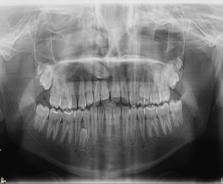
An OPG was taken which showed impacted permanent canine in the first quadrant, bunching of the teeth in the premolar area and supernumerary third molars in the maxillary arch on the both sides. An impacted mandibular premolar was observed in the fourth quadrant.
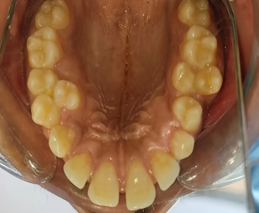
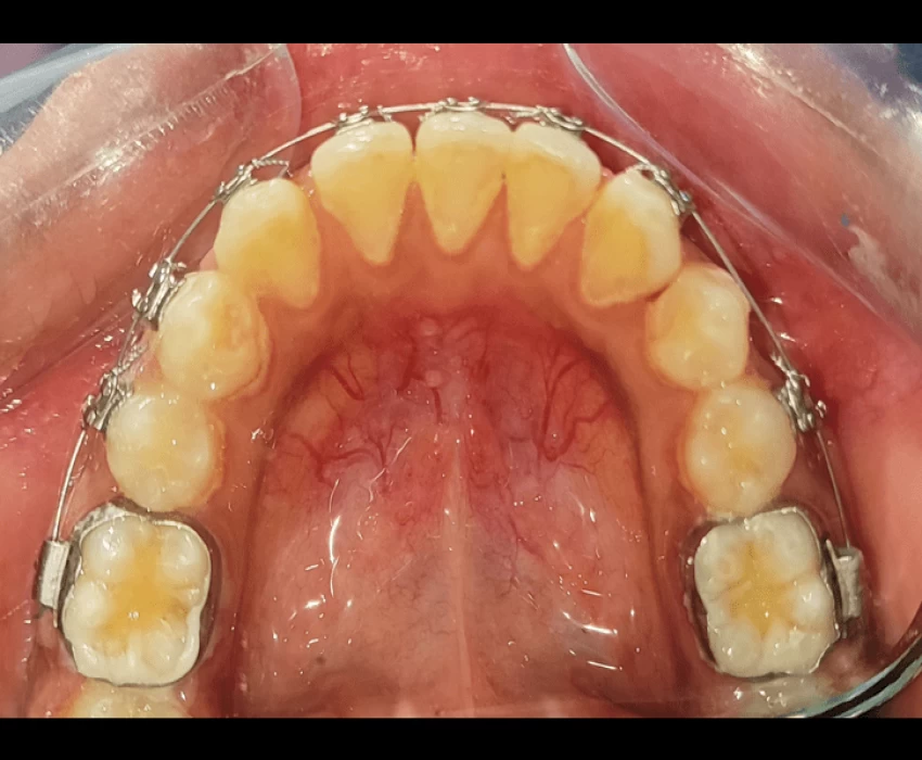
Intraoral examination revealed retained deciduous canine in the first quadrant and supernumerary premolar in the first and second quadrants of the maxilla. Mandibular arch showed missing right permanent lateral incisor.
This is very unusual presentation of supernumerary teeth. As an essential diagnosis aid we usually go for OPG, Lateral cephalogram and PA Cephalogram; which gives us 2D presentation of the hard and soft tissue structures.
Highlights of this case are the unusual number of supernumerary teeth and appearance of hidden supernumerary teeth post extraction of the deciduous canine tooth and Parapremolars.
Case was discussed and after approval of the treatment plan ,the supernumerary premolars in the first and second quadrants of the maxilla along with the retained 53 were extracted.
Patient reported after a gap of one month .It was surprising to see supernumerary canine in the first quadrant and a supernumerary premolar in the second quadrant.
Another OPG was taken at this stage which confirmed the above findings.
References
Sivapathasundharam B. Shafer's Textbook of Oral Pathology-E Book. Elsevier Health Sciences; 2016 Jul 25.
Archana Rani, Arvind Kumar Pankaj, Rakesh Kumar Diwan, Rakesh Kumar Verma, Anita Rani, Jai Prakash Gupta. PREVALENCE OF SUPERNUMERARY TEETH IN NORTH INDIAN POPULATION: A RADIOLOGICAL STUDY. Int J Anat Res 2017;5(2.2):3861-3865.
Mahabob MN, Anbuselvan GJ, Kumar BS, Raja S, Kothari S. Prevalence rate of supernumerary teeth among non-syndromic South Indian population: An analysis. Journal of pharmacy & bioallied sciences. 2012 Aug;4(Suppl 2):S373.
Ata-Ali F, Ata-Ali J, Peñarrocha-Oltra D, Peñarrocha-Diago M. Prevalence, etiology, diagnosis, treatment and complications of supernumerary teeth. Journal of clinical and experimental dentistry. 2014 Oct;6(4):e414.
Mossaz J, Suter VG, Katsaros C, Bornstein MM. Supernumerary teeth in the maxilla and mandible-an interdisciplinary challenge. Part 2: diagnostic pathways and current therapeutic concepts. Swiss dental journal. 2016 Jan 1;126(3):237-59.
Scheiner M, Sampson W. Supernumerary teeth: a review of the literature and four case reports.
Mossaz J, Kloukos D, Pandis N, Suter VG, Katsaros C, Bornstein MM. Morphologic characteristics, location, and associated complications of maxillary and mandibular supernumerary teeth as evaluated using cone beam computed tomography. European journal of orthodontics. 2014 Dec 1;36(6):708-18.
Tumen EC, Yavuz I, Tumen DS, Hamamci N, Berber G, Atakul F, Uysal E. The detailed evaluation of supernumerary teeth with the aid of cone beam computed tomography. Biotechnology & Biotechnological Equipment. 2010 Jan 1;24(2):1886-92.
The case is being treated by Dr.Masum Mohapatra(P.G IIIrd year) under the supervision of Dr.Ragni Tandon(Professor and Head) and Dr.Kamlesh Singh (Professor)


